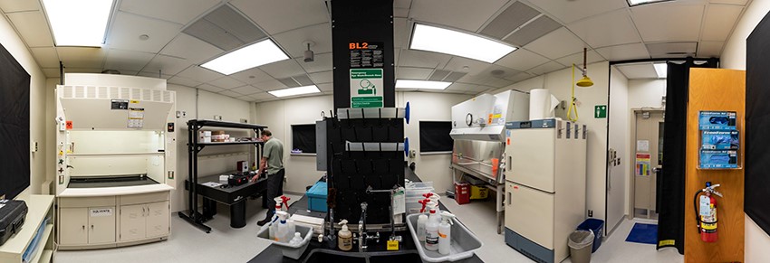
BEAMM Lab explores interactions between energy and tissue
Lincoln Laboratory has a well-established track record of developing advanced imaging technologies. Now, staff are looking to apply that imaging experience to biological research linked to tissues in the new Biophotonic, Electric, Acoustic, and Magnetic Measurement (BEAMM) Lab.
"The BEAMM Lab provides a unique space for rapid, flexible development of novel experimentation related to tissue-technology interactions," said Ryan J. McKindles, who works in the Bioengineering Systems and Technologies Group. "It is a place where we can look at different energy types — such as optical or acoustic energy — and how they interface with tissue."
The BEAMM Lab opened last year on October 1 as a resource that Laboratory groups could use to support their ongoing or future programs. At the facility staff can interrogate cells or tissue, or utilize optical tissue phantoms, which can be produced in any shape and mimic specific material properties of tissue, such as optical or acoustic.
"The BEAMM Lab is a unique resource that will enable multimodal, multispectral characterization of biological materials," said Catherine Cabrera, associate group leader of the Bioengineering Systems and Technologies Group. "Biomedical imaging has been identified as a key area in which the Laboratory could have a significant impact by leveraging a range of resources, expertise, and capabilities in optics, lasers, radiofrequency, images, prototyping, image processing, and data analysis. The BEAMM Lab will create a collaborative space in which this vision can come closer to reality."
The 320-square-foot facility offers access to a biosafety cabinet, a chemical fume hood, an optical table and appropriate refrigeration, safety equipment, and supplies for handling biological materials. It also is a Biosafety Level 2 facility, which means it can support the study of human tissue.

Researchers studying advanced imager technology and bioengineering systems, as well as those at the Lincoln Laboratory Supercomputing Center (LLSC), are currently using the BEAMM Lab for research in two programs. The first program utilizes a novel type of hyperspectral microscope to scan tissue and investigate what is inside without destroying it. The second program explores how direct energy — such as lasers or RF energy — interacts with tissue.
"These [types of] experiments could further our understanding of basic physiology, assist in the research and development of novel sensing and stimulation devices, and support rapid physiological modeling at a tissue and cellular level," McKindles said.
Staff are looking to broaden their reach across several biological areas, from cells to cognition. For example, one current application that uses deep learning to predict standard tissue stains from the hyperspectral images would eliminate the need for volatile chemicals in the field. Additionally, the application of novel tissue-modeling methods could improve the scientific understanding of medical conditions such as cancer and Alzheimer’s disease. In the future, staff also plan to install a faster network connection to the LLSC to enable real-time data acquisition and modeling for adaptive designs.
"What I find most exciting about the BEAMM Lab," Cabrera said, "is that it offers a multidisciplinary space for Laboratory staff to work together on amazing research and development projects that really couldn’t be done anywhere else."